THE EYE
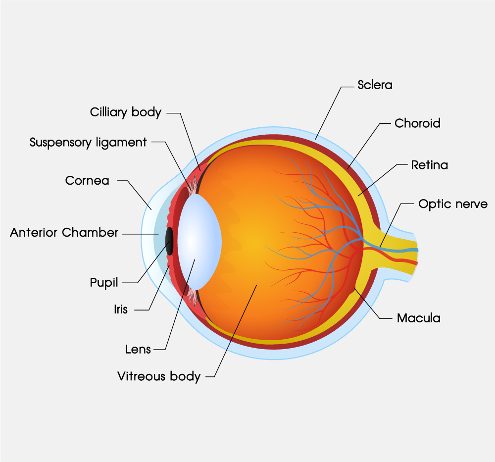
Cornea
The cornea is the clear, transparent part located at the front of the eye structure. It is the primary refractive surface and it helps focus light into the eye.
Iris
The iris is the colored part of the eye surrounded by muscles that help regulate the amount of light that enters the eye. The pupil is the black center of the iris. It appears black because light passing through the eye is absorbed by tissues.
Crystalline lens
Located inside the eyeball and behind the iris, the crystalline lens is a transparent structure that focuses light rays onto the retina. The optical lens adjusts by changing its shape to help the eye focus at different distances.
Retina
The retina is the nerve layer lining the back of the eye. It contains millions of light receptors (also known as photoreceptors). The retina senses light focused by the cornea and the crystalline lens and sends impulses through the optic nerve to the brain.
Read More
AQUEOUS HUMOR
The eye’s spherical shape is maintained by “aqueous humor”, a transparent, jelly-like substance that fills about 2/3 of the space within the eye and is located between the crystalline lens and the retina.
Macula
The macula is part of the retina, approximately 5.5 mm, has the highest density of photoreceptors, and is free of major blood vessels. The fovea sits in the center and is responsible for sharp and detailed vision.
Choroid
The choroid is known as the vascular layer of the eye and it provides oxygen and nourishment to the retina.
Sclera
The sclera, also known as the white of the eye, is the protective outer layer of the eye.
Ciliary body
The ciliary body is a part of the eye that comprises the ciliary muscle. It lies behind the iris and is attached to the lens by connective tissue called the zonular fibers. Relaxation of the ciliary muscle puts tension on these fibers. As a result, it changes the lens shape to control the focus of light on the retina at various distances. This process is called accommodation.
THE FUNCTIONS OF EYE MUSCLES
Our eyes have to move continually to follow a target because only a tiny part of the eye, the fovea, provides sharp vision. Each eye’s movement is controlled by six small muscles that allow us to look up, down, left, right, and for the eye to rotate in and out.
These eye muscles handle a heavy load as they carry out three actions simultaneously: focusing, converging, and accommodating for clear vision and field depth. As a result, the lens changes its shape, the pupil changes its size, and the eyes rotate to keep focus.
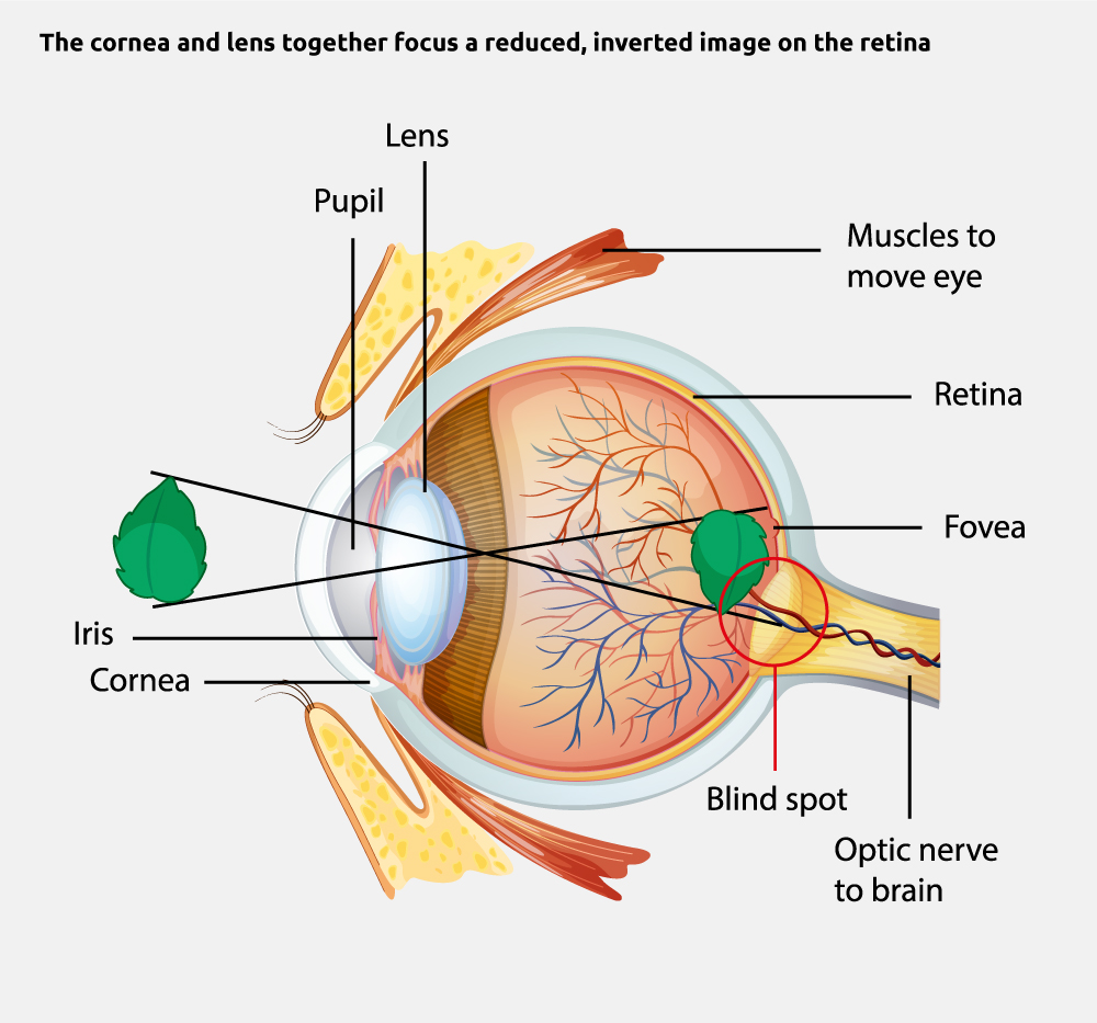
VISION
HOW OUR EYES FORM AN IMAGE
Light enters the eye through the cornea, which acts as a convex lens and does most of the refracting in the eye. The light then continues through the pupil, which controls how much light enters and passes through the eye. The lens is convex on both sides. It contracts and relaxes to control the amount of refraction and move the focal point to make fine adjustments for near and far objects.
When light passes through the clear center of the eye, it strikes the retina, which contains specialized cells that respond to light. Some of these cells then send signals through the optic nerve to the brain, which interprets these signals as images.